What Is Vascular Bundle And Its Types Best Design Idea
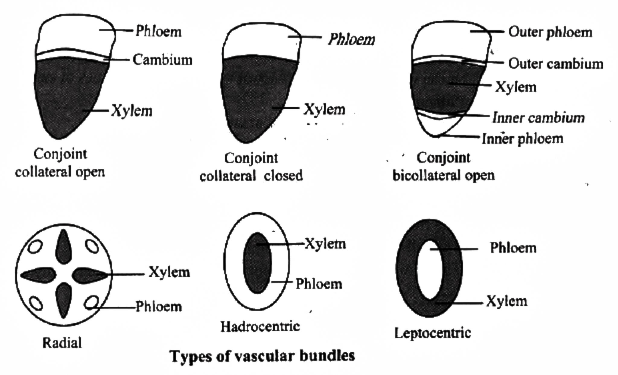
A group of xylem and phloem forms a vascular bundle. The different types of vascular bundles met within plants are: 1. Radial bundles (Simple): Xylem and phloem are seen as patches and they alternate each other, and occupy the different radii on the axis separated by non conductive tissue. example: Dicot and monocot roots. 2.
class 11th Anatomical structure of root and Vascular tissue system 02 06 2020 YouTube
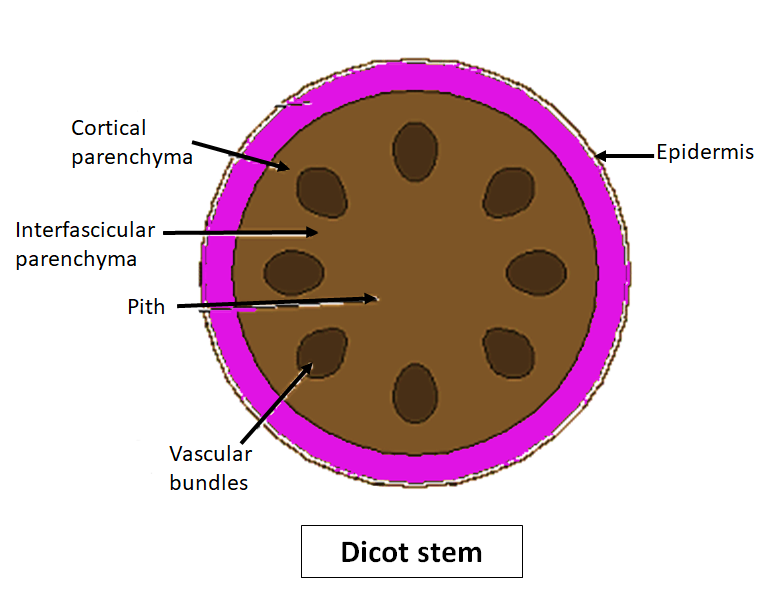
Vascular bundle; Answer . The diagram shows a cross section of a dicot stem, and we need to identify one of the structures in it. To do this, let's look at the different structures in a dicot stem and their functions. The epidermis is a single layer of cells that forms the outer covering of the plant stem..
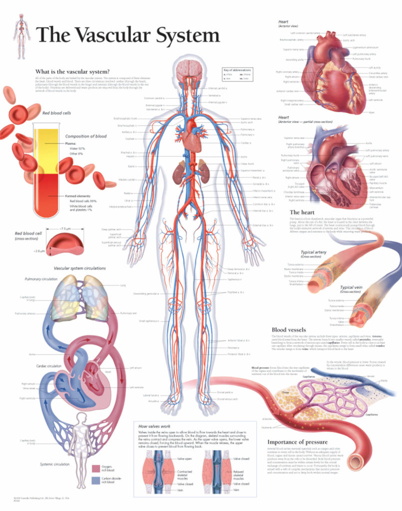
The Vascular System Scientific Publishing
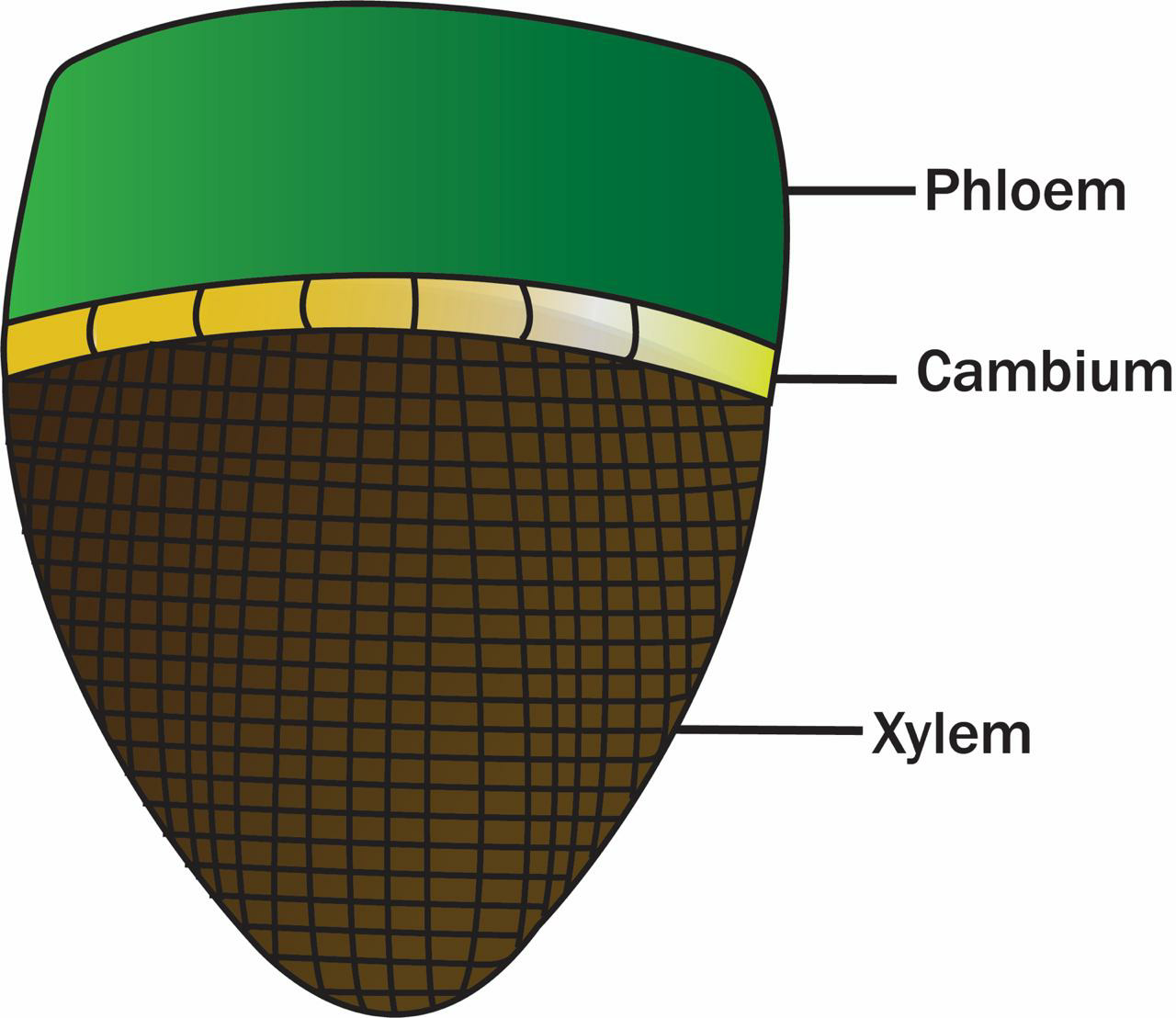
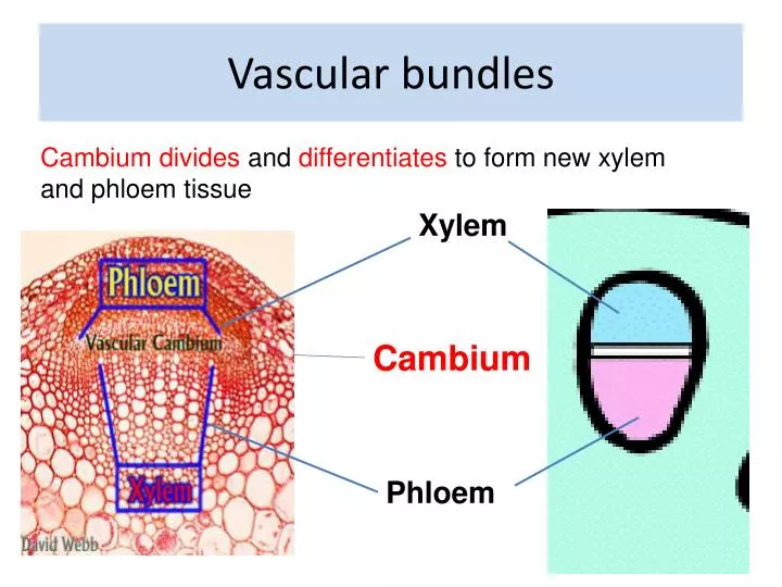
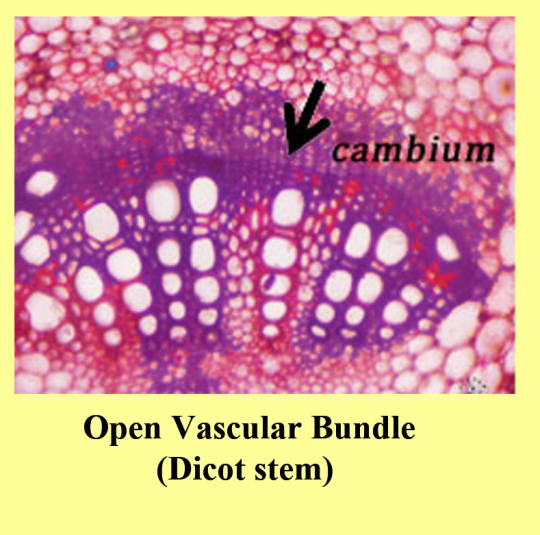
phloem together constitute vascular bundles (Figure 6.2). In dicotyledonous stems, cambium is present between phloem and xylem. Such vascular bundles because of the presence of cambium possess the ability to form secondary xylem and phloem tissues, and hence are called open vascular bundles. In the monocotyledons, the vascular bundles have no
PPT Roots PowerPoint Presentation, free download ID4007333
1. Vascular bundle contains a strip of cambium in between phloem and xylem. 2. Phloem and xylem do not lie in direct contact with each other. ADVERTISEMENTS: 3. Due to activity of cambium, original or primary phloem and xylem move away from each other. Secondary phloem and secondary xylem are formed in between. 4.
What Is Vascular Bundle Hindi Best Design Idea
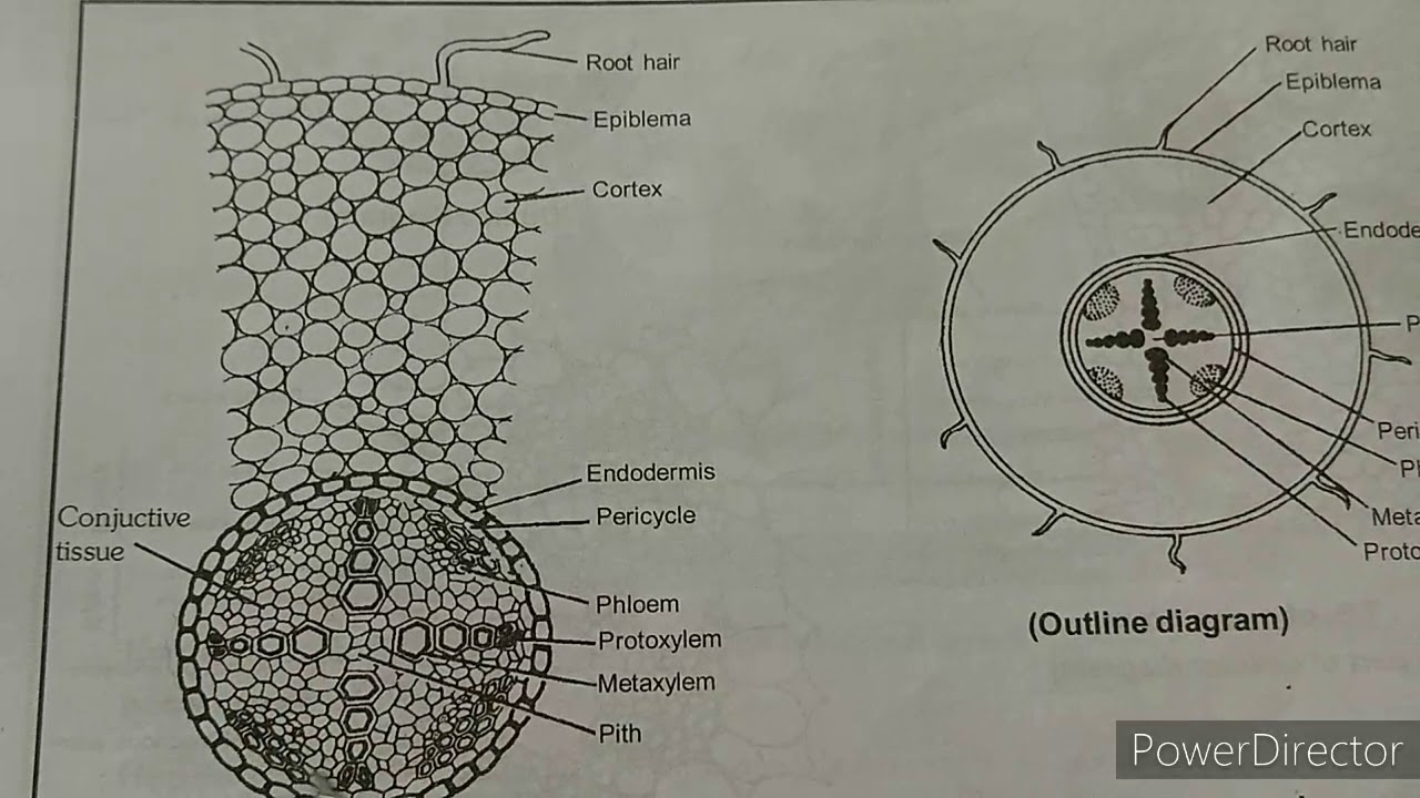
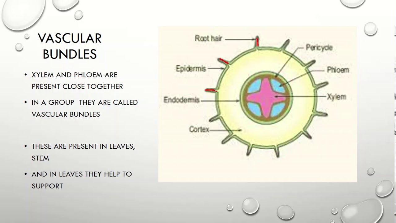
a). Hypodermis b). Outer cortex c). Inner cortex d). Endodermis (3). Stele a). Pericycle b). Vascular bundles c). Medullary rays d). Pith (1). Epidermis Ø Epidermis is the outermost layer, composed of parenchymatous cells. Ø Usually, epidermis composed of single layer of cells. Ø Cells are closely packed without any intercellular spaces.
PPT Vascular bundles PowerPoint Presentation, free download ID1710731
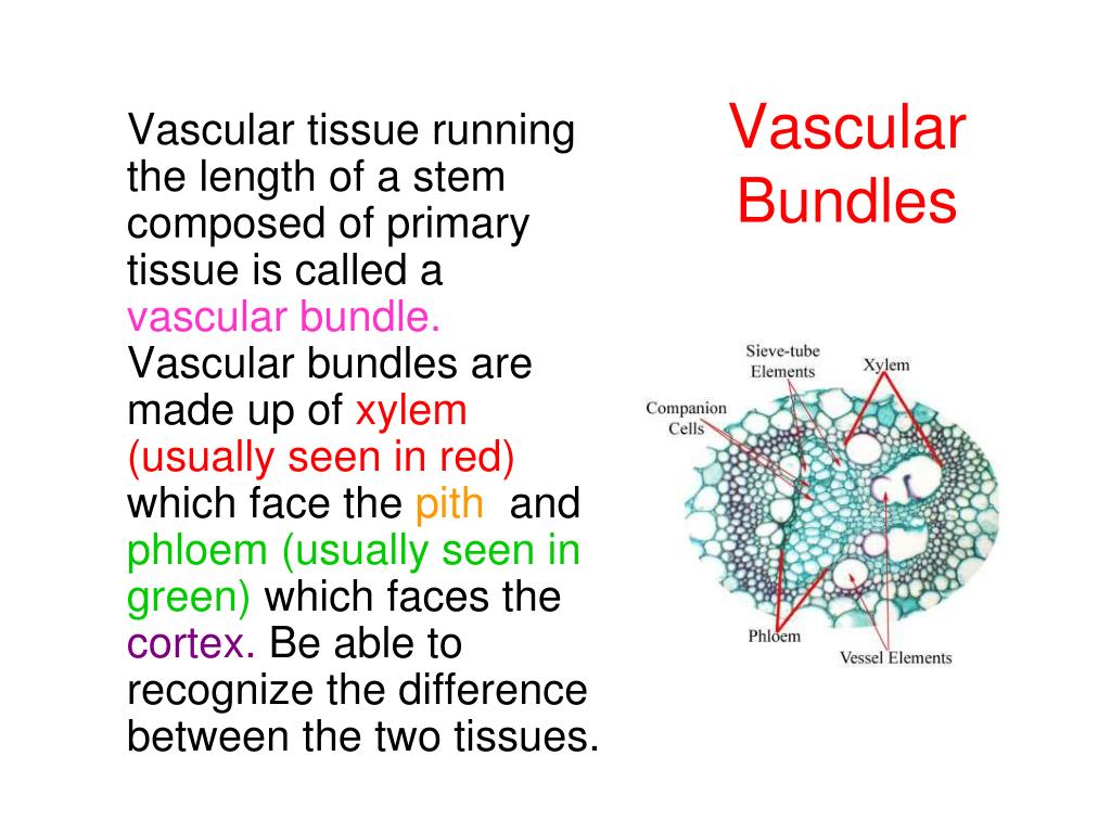
A vascular bundle is a part of the transport system in vascular plants. The transport itself happens in the stem, which exists in two forms: xylem and phloem. Both these tissues are present in a vascular bundle, which in addition will include supporting and protective tissues.
Department of Botany
There are open vascular bundles when a vascular cambium layer (in dicots) is present. Those devoid of it (in monocots) are closed vascular bundles. Thus, vascular bundles typically represent the organization of xylem and phloem and their association with other accessory transporting tissues.
Monocot Stem Labeled
Type # 1. Collateral Bundle: A vascular bundle in which a strand c f phloem is present external to the strand of xylem on the same radius side by side is known as collateral bundle. ADVERTISEMENTS: Cambium may be present or absent in between xylem and phloem, and so there are the following two types of collateral bundle:

Identify the vascular bundles given in the following figures . Sarthaks eConnect Largest
Since there is no ring of vascular bundles, there is no "inside" pith and "outside" cortex. All the ground tissue is considered to be cortex. Monocot corn stem cross section showing vascular bundles. Melissa Ha. CC BY-NC 2.0. In monocot vascular bundles the phloem is always oriented toward the outside of the plant and the xylem toward.

Anatomy of the vascular bundles. LM images of cross sections taken... Download Scientific Diagram
In monocot stems, the vascular bundles are randomly scattered throughout the ground tissue (Figure 30.9). Figure 30.9 In (a) dicot stems, vascular bundles are arranged around the periphery of the ground tissue. The xylem tissue is located toward the interior of the vascular bundle, and phloem is located toward the exterior. Sclerenchyma fibers.

Stem Growth Biology for Majors II
Solution Verified by Toppr An open vascular bundle is characterized by the presence of cambial ring between the xylem and phloem bundles. These are seen in dicot stem which undergoes secondary. The diagram represents the open vascular bundle. Solve any question of Anatomy Of Flowering Plants with:- Patterns of problems > Was this answer helpful? 0
Radial vascular bundles occur inA. StemB. Monocot rootC. Dicot rootD. Both monocot and dicot roots
As leaf traces connect the vascular bundle system of the trunk with the vascular bundles of the petioles, cross-sections from the cortical zone of the trunk and from a petiole were also stained (Fig. 7b and c). In leaf traces of the outer cortical zone of the trunk, a second fibre cap seemed to develop on the xylem side of the vascular bundles as fibre cells were seen on this side, opposite to.
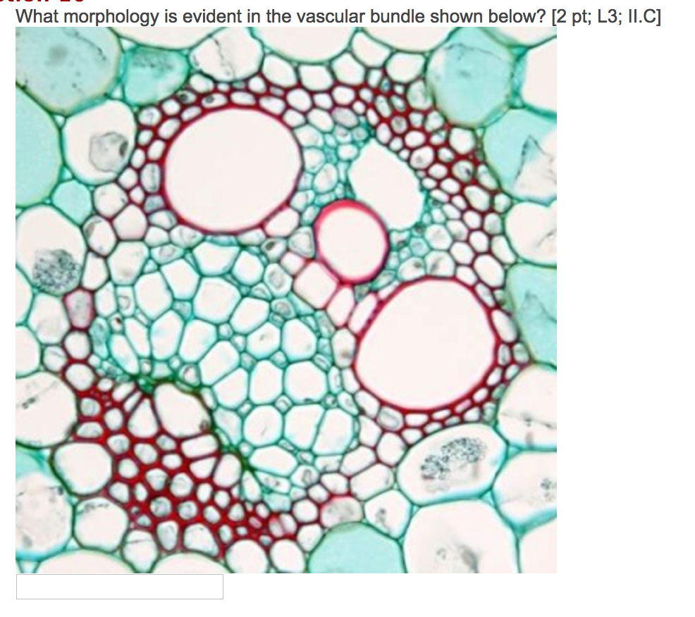
Solved What morphology is evident the vascular bundle shown
Image by RolfDieterMueller ( CC-BY) Figure 3.3.2.3 3.3.2. 3: In eusteles (left), vascular bundles are arranged around the periphery of the ground tissue. The xylem tissue is located toward the interior of the vascular bundle, and phloem is located toward the exterior. Primary phloem fibers cap the vascular bundles.
How open vascular bundles differ from closed vascular bundles?
Bamboos have a hierarchical gradient structure, that is, a macroscopic gradient structure in culm diameter and a microscopic one in the bundle sheath distribution [1-4].A bamboo culm is made up of two kinds of cells, matrix tissue cells (parenchymatous) and sclerenchyma cells (vascular bundle) [5, 6].Vascular bundles made up of sclerenchyma cells act as reinforcement in bamboo.
Vascular bundles in root and stem YouTube
Download scientific diagram | Schematic diagram for measuring the radial length/tangential diameter of an open vascular bundle in F. yunnanensis culm. M: metaxylem, P: phloem, Px: protoxylem, R.
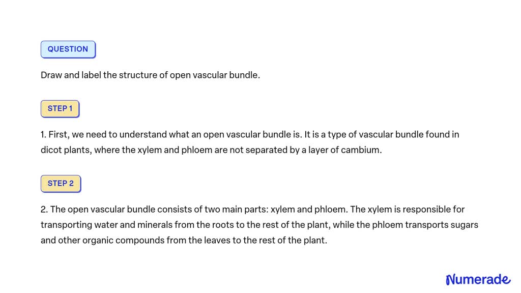
SOLVED Draw and label the structure of open vascular bundle.
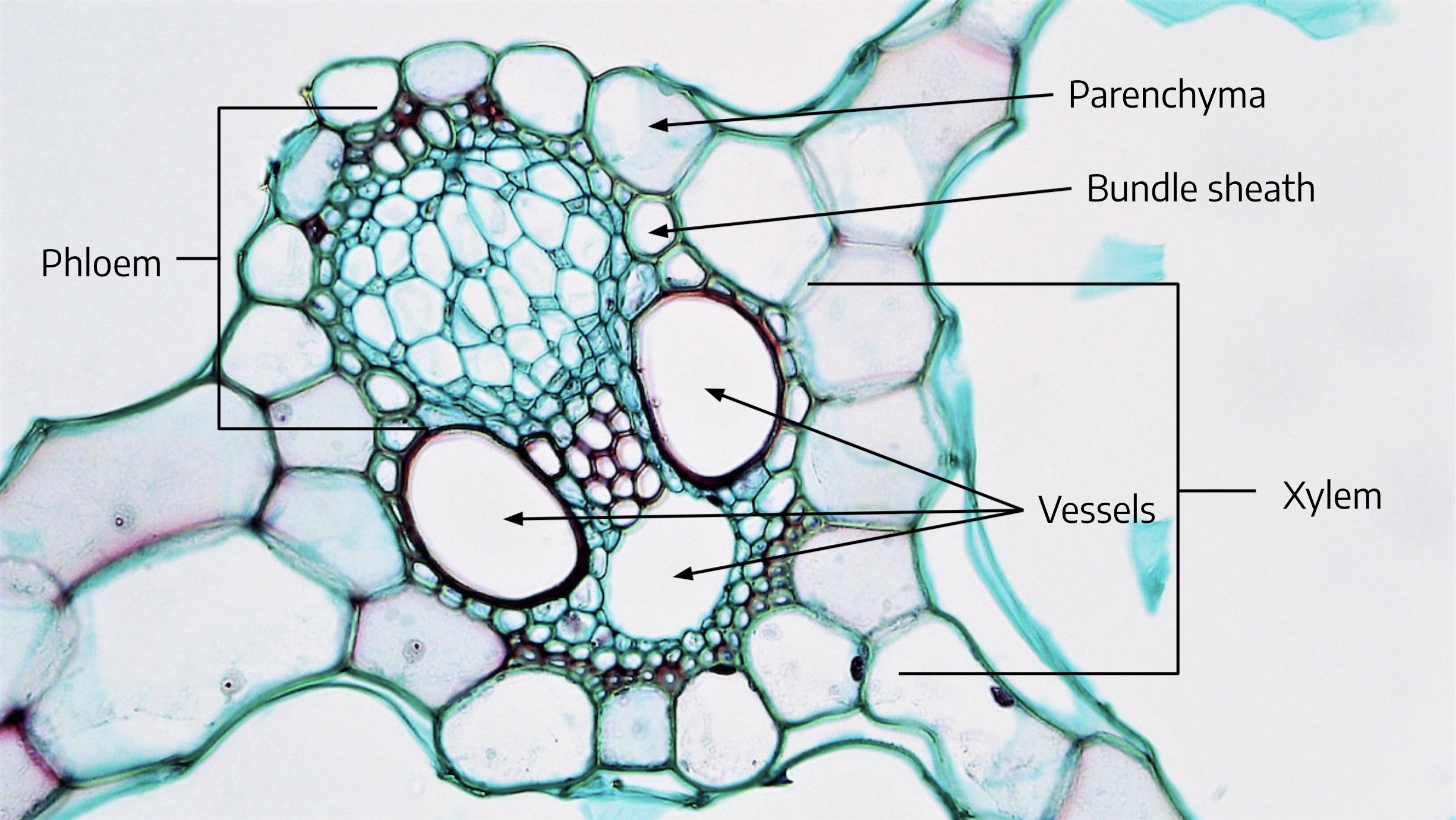
In the larger vascular bundle, it is easier to distinguish the large, open vessel elements (stained red). Within the vascular bundle, the xylem tissue is closer to the upper epidermis and the phloem tissue is closer to the lower. Each vascular bundle is surrounded by larger cells with darkly-stained contents. These make up the bundle sheath.
